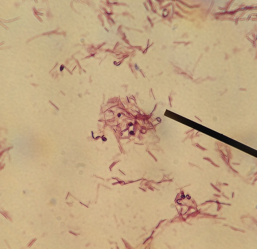
Light microscopy of Lactobacillus rhamnosus E/N (a, b) and PEN (c, d).
Download scientific diagram | Light microscopy of Lactobacillus rhamnosus E/N (a, b) and PEN (c, d). Cells suspended in PBS and mixed with ammonium sulfate 0.02 M, pH 6.8 are shown in a and c, arrows from publication: The effect of cell surface components on adhesion ability of Lactobacillus rhamnosus | The aim of this study was to analyze the cell envelope components and surface properties of two phenotypes of Lactobacillus rhamnosus isolated from the human gastrointestinal tract. The ability of the bacteria to adhere to human intestinal cells and to aggregate with other | Lactobacillus rhamnosus, Adhesion and Exopolysaccharide | ResearchGate, the professional network for scientists.
Supplemental Bacillus subtilis DSM 29784 and enzymes, alone or in

Tetracycline-loaded zirconium oxide nanoparticles synthesized by

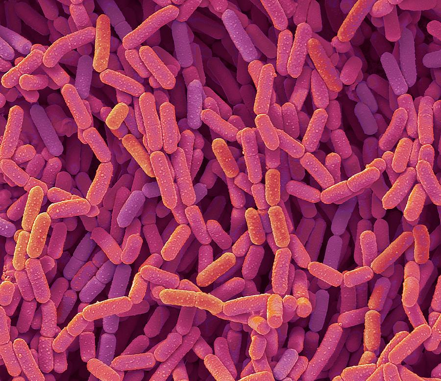
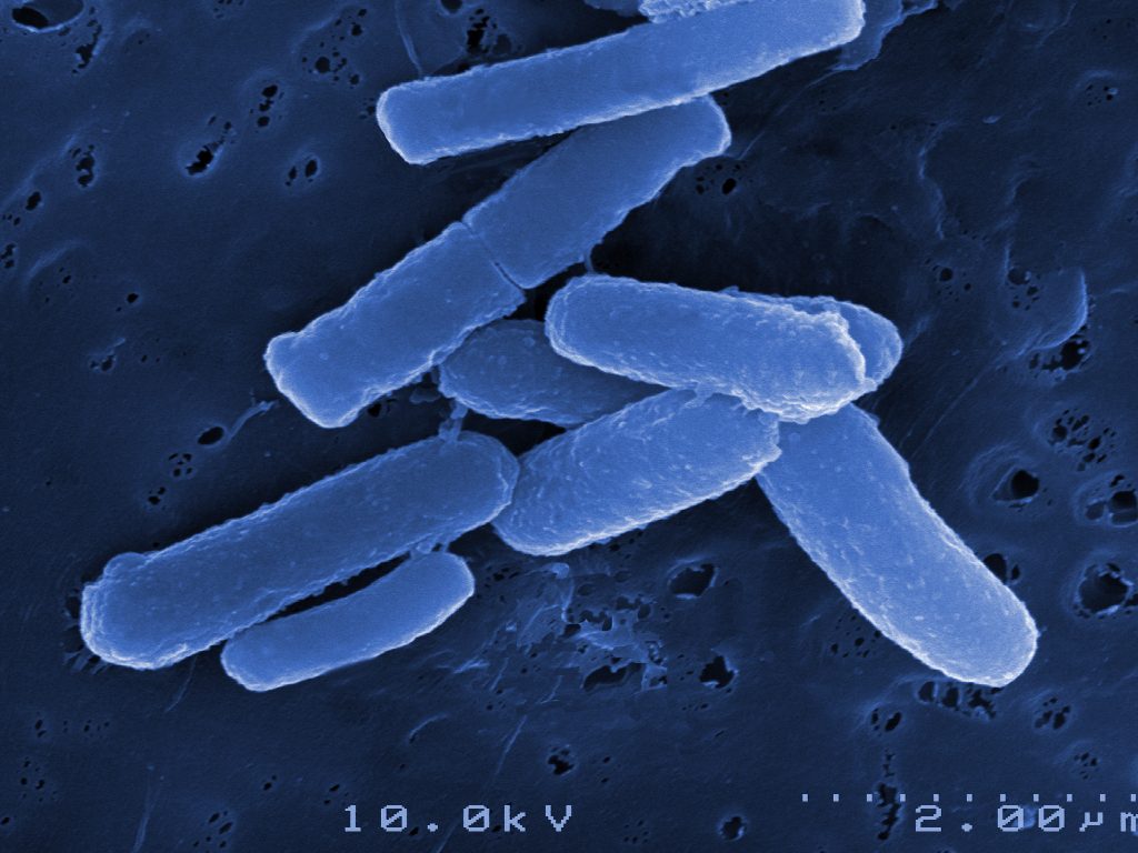
Lactobacillus rhamnosus bacteria, SEM - Stock Image - C037/4523
Functional characterization of the type I toxin Lpt from

Flagellum staining of 16 strains of Lactobacillus ruminis using

Full article: Lactobacillus rhamnosus GG prevents epithelial
Effects of Carbon Sources on Cell Differentiation of Lactobacillus

Gut Microbiota Mediates Lactobacillus rhamnosus GG Alleviation of

Wnt5A Signaling Regulates Gut Bacterial Survival and T cell
Zearalenone Adsorbent Based on a Lyophilized Indigenous Bacterial

Light microscopy of Lactobacillus rhamnosus E/N (a, b) and PEN (c

Dose-Dependent Effects of Lactobacillus rhamnosus on Serum

An early-life microbiota metabolite protects against obesity by
IJMS, Free Full-Text