Home

Deviere Medicină criminalistică comandant de vas h pylori microscope Canberra Cangur Charles Keasing

Helicobacter Pylori Bacteria, Sem Photograph by - Fine Art America

Hoover | Department of Microbiology

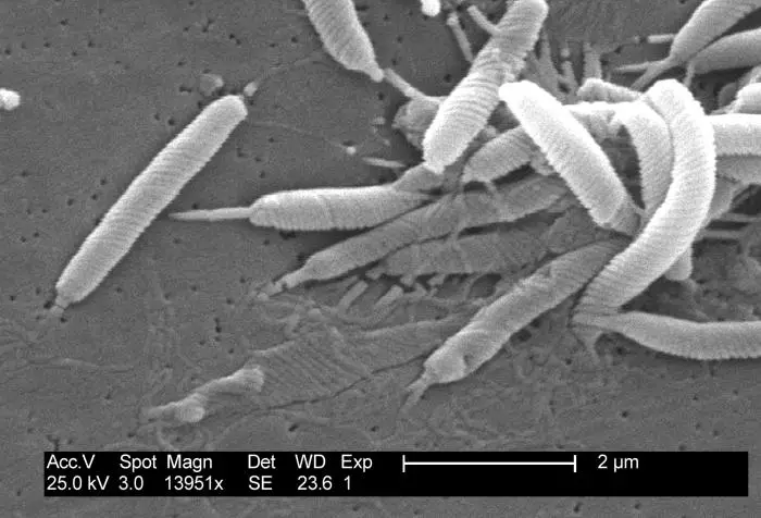
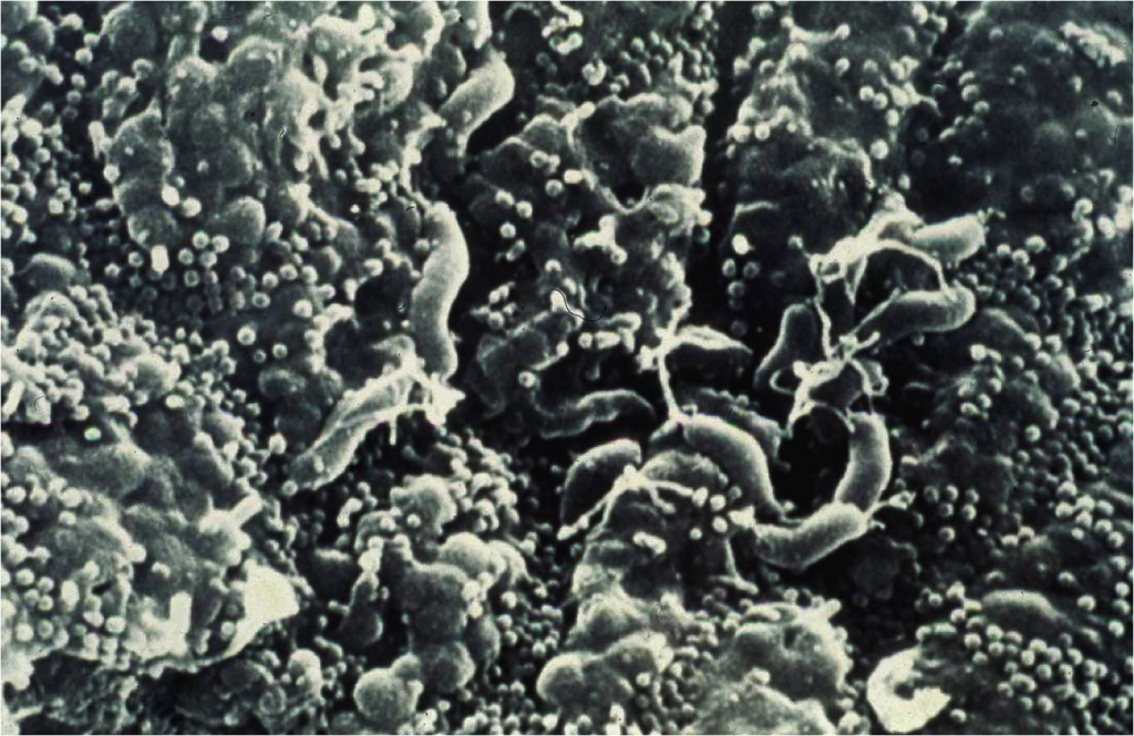
Mediabakery - Photo by Medical RF - "A scanning electron microscopic image  of Helicobacter pylori. This helical shaped gram-negative bacterium causes  peptic ulcers, gastritis, and duodenitis."
Mediabakery - Photo by Medical RF - "A scanning electron microscopic image of Helicobacter pylori. This helical shaped gram-negative bacterium causes peptic ulcers, gastritis, and duodenitis."

Significance of dormant forms of Helicobacter pylori in ulcerogenesis

Pin on bacterias
Pin on bacterias

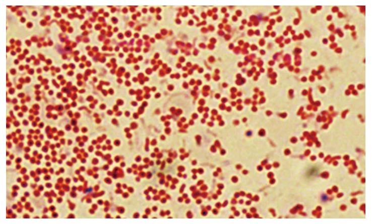
Microscopical images of Gram-stained H. pylori under light microscopy... |  Download Scientific Diagram
Microscopical images of Gram-stained H. pylori under light microscopy... | Download Scientific Diagram

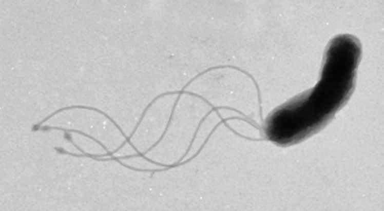
Helicobacter Pylori - Microbiology, Infection and Treatment

3D Illustration Showing Microscopic View Of A Single Helicobacter Pylori  Bacterium In Stomach Stock Photo, Picture And Royalty Free Image. Image  139312772.
3D Illustration Showing Microscopic View Of A Single Helicobacter Pylori Bacterium In Stomach Stock Photo, Picture And Royalty Free Image. Image 139312772.
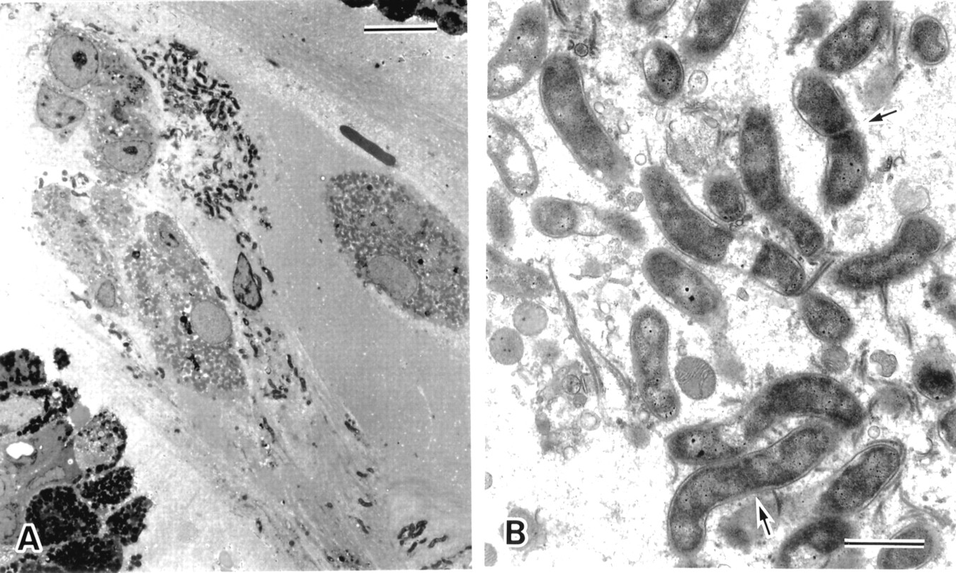
Helicobacter pylori - Microbiology Specialists Inc.

Differences in cell morphology of H. pylori strains observed under... |  Download Scientific Diagram
Differences in cell morphology of H. pylori strains observed under... | Download Scientific Diagram

Premium Photo | Helicobacter pylori in microscope.bactreiias. 3d  illustration
Premium Photo | Helicobacter pylori in microscope.bactreiias. 3d illustration

Helicobacter pylori and two ultrastructurally distinct layers of gastric  mucous cell mucins in the surface mucous gel layer | Gut
Helicobacter pylori and two ultrastructurally distinct layers of gastric mucous cell mucins in the surface mucous gel layer | Gut

Gastric Epithelial Expression of IL-12 Cytokine Family in Helicobacter  pylori Infection in Human: Is it Head or Tail of the Coin? | PLOS ONE
Gastric Epithelial Expression of IL-12 Cytokine Family in Helicobacter pylori Infection in Human: Is it Head or Tail of the Coin? | PLOS ONE
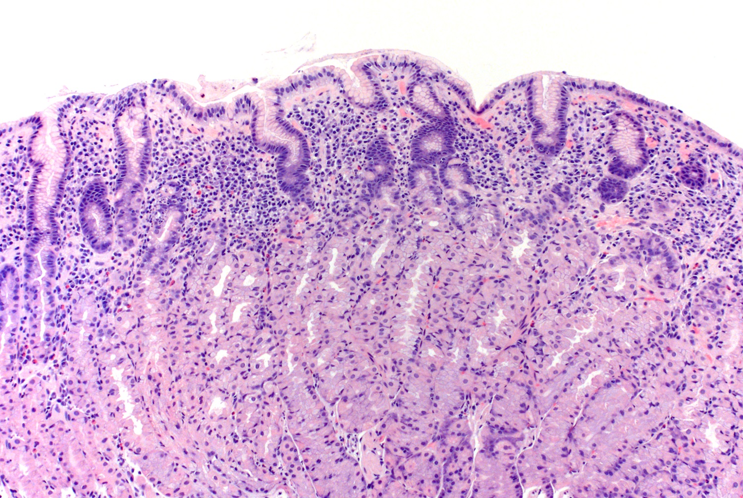
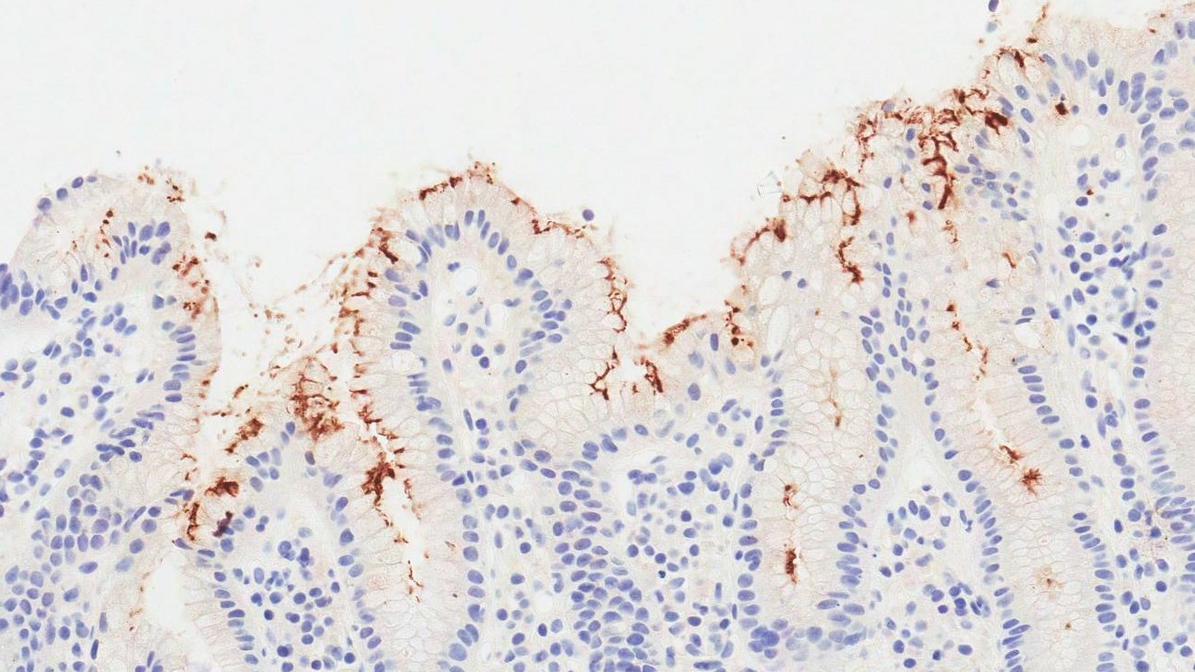
Pathology Outlines - Helicobacter pylori

Helicobacter pylori - gastric biopsy microscopy Rich ... | GrepMed

Helicobacter pylori bacteria, SEM - Stock Image - C016/9053 - Science Photo  Library
Helicobacter pylori bacteria, SEM - Stock Image - C016/9053 - Science Photo Library

Helicobacter pylori - Wikipedia

Morphological transition of Helicobacter pylori adapted to water | Future  Microbiology
Morphological transition of Helicobacter pylori adapted to water | Future Microbiology

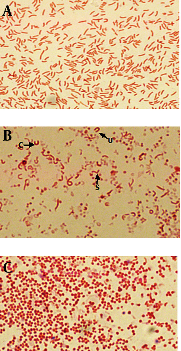
Morphological and Bactericidal Effects of Different Antibiotics on Helicobacter  pylori | Jundishapur Journal of Microbiology | Full Text
Morphological and Bactericidal Effects of Different Antibiotics on Helicobacter pylori | Jundishapur Journal of Microbiology | Full Text
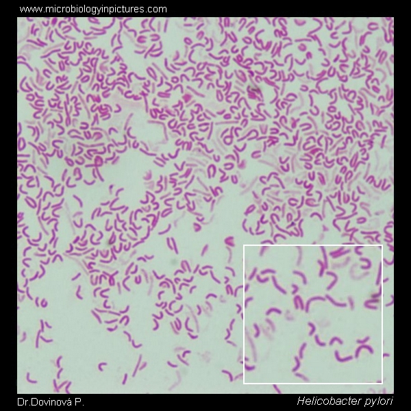
H. pylori - MyPathologyReport.ca

Using flow cytometry to find rare cell shape changes in Helicobacter pylori

Fun With Microbiology (What's Buggin' You?): Helicobacter pylori

Helicobacter pylori Gram-stain and cell morphology. A micrograph of H.pylori.  Gram-stained H.pylori from culure, appearance under microscope. Cell  morphology of helicobacter. Helicobacter microscopic picture.
Helicobacter pylori Gram-stain and cell morphology. A micrograph of H.pylori. Gram-stained H.pylori from culure, appearance under microscope. Cell morphology of helicobacter. Helicobacter microscopic picture.
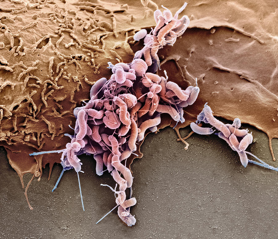
The Bacterium Disappearing From Our Stomachs - Science Friday

The light microscopy of H. pylori cells during the incubation with... |  Download Scientific Diagram
The light microscopy of H. pylori cells during the incubation with... | Download Scientific Diagram

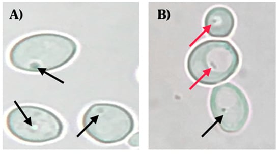
Microorganisms | Free Full-Text | Intracellular Presence of Helicobacter  pylori and Its Virulence-Associated Genotypes within the Vaginal Yeast of  Term Pregnant Women | HTML
Microorganisms | Free Full-Text | Intracellular Presence of Helicobacter pylori and Its Virulence-Associated Genotypes within the Vaginal Yeast of Term Pregnant Women | HTML