Patient 3. Axial Spin Echo T1-weighted (A), Turbo Spin Echo T2-weighted
Download scientific diagram | Patient 3. Axial Spin Echo T1-weighted (A), Turbo Spin Echo T2-weighted (B), susceptibility weighted (C), diffusion weighted (b1000 and ADC map, D,E) and Turbo Field Echo T1-weighted after contrast administration (F) images. A left basal ganglia lesion also involving thalamus, corona radiata, deep temporal white matter and corpus callosum is appreciated. The lesion showed inhomogeneous signal intensity, with cystic and hemorrhagic components, and moderate enhancement. from publication: Germ cell tumors in male patients without gonadal involvement: Computed tomography/magnetic resonance imaging findings and diagnostic workflow | Germ Cell Tumors, Germ Cell and Embryonal Neoplasms and Gonads | ResearchGate, the professional network for scientists.

Frontiers Turbo Gradient and Spin Echo PROPELLER-Diffusion Weighted Imaging for Orbital Tumors: A Comparative Study With Readout-Segmented Echo-Planar Imaging

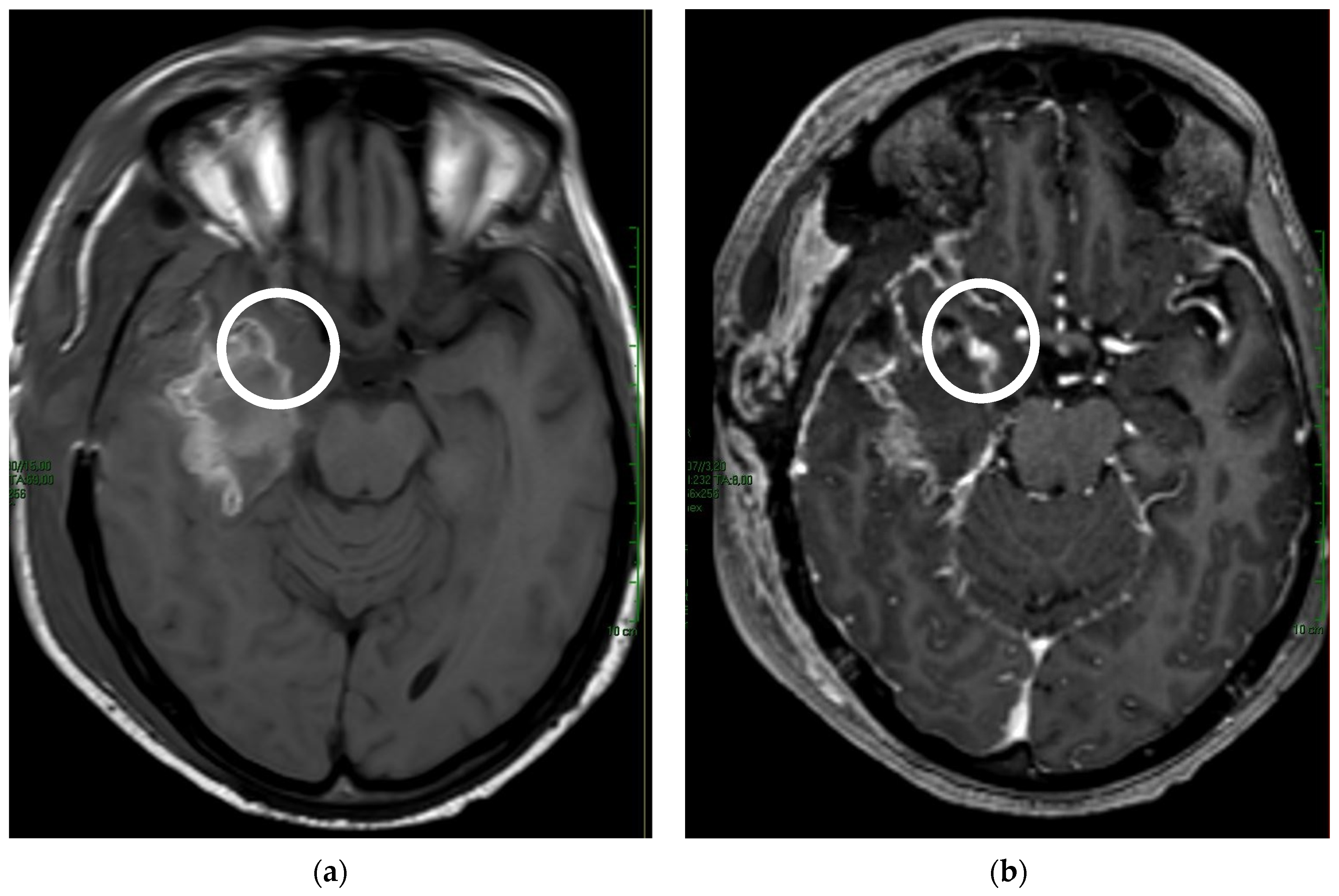
Axial T1-weighted spin-echo (SE)(TR/TE; 500/15 ms)(A, B) and

Introduction to MRI: Basic Pulse Sequences, TR, TE, T1 and T2 weighting

Prospective Comparison of Standard and Deep Learning–reconstructed Turbo Spin-Echo MRI of the Shoulder

MRI sequences: fast spin echo

Comparison of 2D single-shot turbo-spin-echo and spin-echo echo-planar diffusion weighted brain MRI at 3.0 Tesla: preliminary experience in children - ScienceDirect
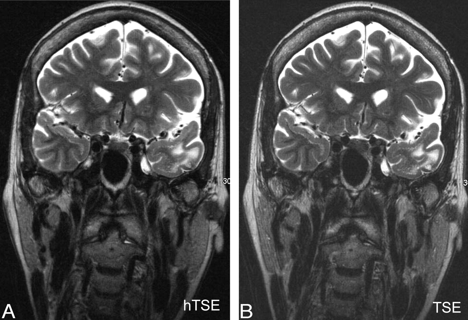
A-D) Brain MRI. Axial turbo spin-echo (TSE) T2-weighted image (A) well
Tomography, Free Full-Text

T1 MRI MRI T1 weighted sequences
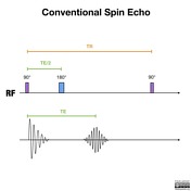
Spin echo sequences, Radiology Reference Article
Fast Spin Echo Sequence: MRI Sequences Guide