Nerf accessoire - e-Anatomy - IMAIOS
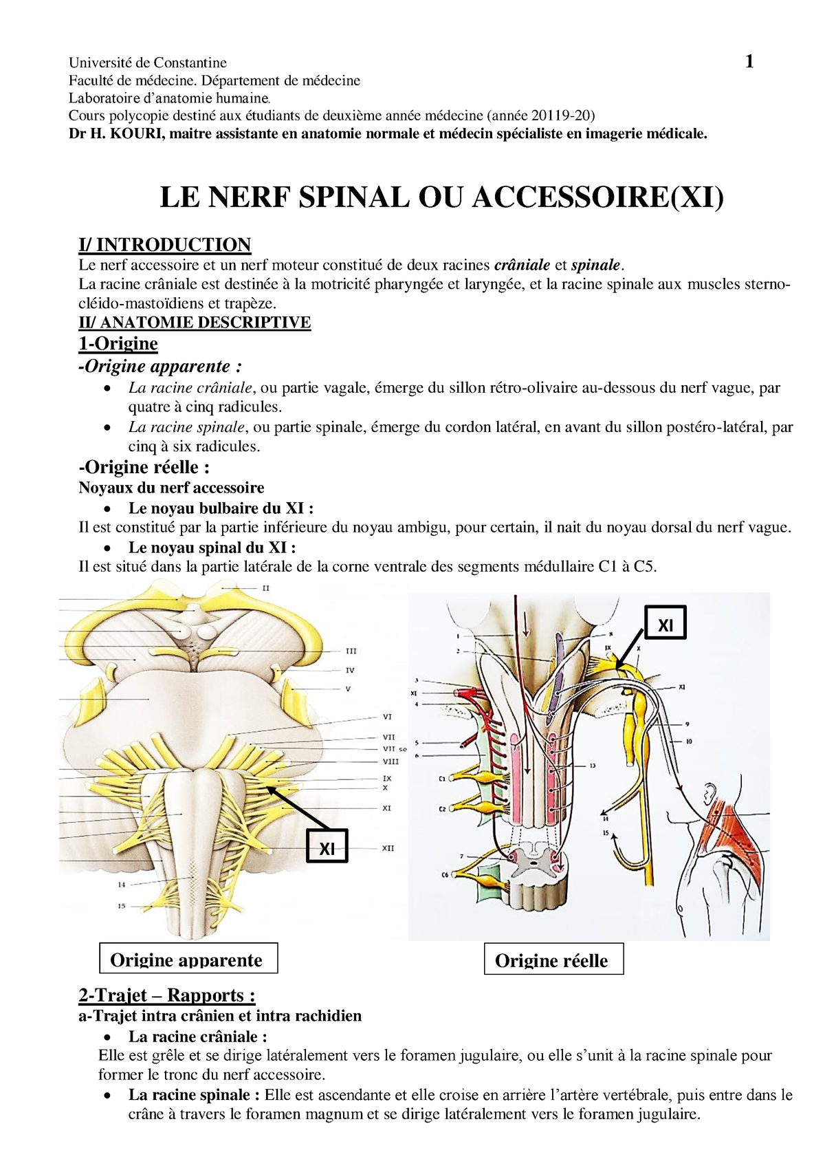
Number: XI Name: Accessory (often separated into thecranial accessoryand spinal accessorynerves) Sensory, motor, or both: Mainly motor Origin/Target: Cranial and Spinal Roots Nuclei: Nucleus ambiguus, Spinal accessory nucleus Function: Controls the sternocleidomastoid and trapezius muscles, and overlaps with functions of the vagus nerve (CN X). Symptoms of damage: inability to shrug, weak head movement. Located in the jugular foramen. Description: The accessory nerve consists of two parts: a cranial and a spinal. The Cranial Part (ramus internus; accessory portion) is the smaller of the two. Its fibersarise from the cells of the nucleus ambiguus and emerge as four or five delicate rootlets from the side of the medulla oblongata, below the roots of the vagus. It runs lateralward to the jugular foramen, where it interchanges fibers with the spinal portion or becomes united to it for a short distance; here it is also connected by one or two filaments with the jugular ganglion of the vagus. It then passes through the jugular foramen, separates from the spinal portion and is continued over the surface of the ganglion nodosum of the vagus, to the surface of which it is adherent, and is distributed principally to the pharyngeal and superior laryngeal branches of the vagus. Through the pharyngeal branch it probably supplies the Musculus uvulæ and Levator veli palatini. Some few filaments from it are continued into the trunk of the vagus below the ganglion, to be distributed with the recurrent nerve and probably also with the cardiac nerves.The Spinal Part (ramus externus; spinal portion) is firm in texture, and its fibers arisefrom the motor cells in the lateral part of the anterior column of the gray substance of the medulla spinalis as low as the fifth cervical nerve. Passing through the lateral funiculus of the medulla spinalis, they emerge on its surface and unite to form a single trunk, which ascends between the ligamentum denticulatum and the posterior roots of the spinal nerves; enters the skull through the foramen magnum, and is then directed to the jugular foramen, through which it passes, lying in the same sheath of dura mater as the vagus, but separated from it by a fold of the arachnoid. In the jugular foramen, it receives one or two filaments from the cranial part of the nerve, or else joins it for a short distance and then separates fromit again. As its exit from the jugular foramen, it runs backward in front of the internal jugular vein in 66.6 per cent. of cases, and behind in it 33.3 per cent. (Tandler). The nerve then descends obliquely behind the Digastricus and Stylohyoideus to the upper part of the Sternocleidomastoideus; it pierces this muscle, and courses obliquely across the posterior triangle of the neck, to end in the deep surface of the Trapezius. As it traverses the Sternocleidomastoideus it gives several filaments to the muscle, and joins with branches from the second cervical nerve. In the posterior triangle it unites with the second and third cervical nerves, while beneath the Trapezius it forms a plexus with the third and fourth cervical nerves, and from this plexus fibers are distributed to the muscle.

Nerf mandibulaire - e-Anatomy - IMAIOS

Nerf vague - e-Anatomy - IMAIOS

Nerf vague - e-Anatomy - IMAIOS

Nerf accessoire - e-Anatomy - IMAIOS

Nerf obturateur accessoire - e-Anatomy - IMAIOS

Nerf accessoire - e-Anatomy - IMAIOS

Nerf optique - e-Anatomy - IMAIOS

E-Anatomy Premium 4.12.6 – اپلیکیشن آموزش کامل آناتومی بدن اندروید

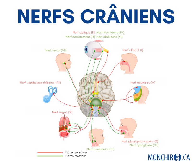
Nerfs crâniens - e-Anatomy - IMAIOS

Accessory nerve - e-Anatomy - IMAIOS

حمّل مجانًا e-Anatomy حزمة تطبيق أندرويد الخاصة بنظام الأندرويد
Protect your bones and joints

Nerf spinal - e-Anatomy - IMAIOS