Home

reputatie cred că sunt bolnav Timpul zilei cell under electron microscope Fobie plin de speranță montură

2.3.3 Identify structures from electron micrographs of liver cells - YouTube

Frontal Cortex — Making scientific images : Electron microscopy (1)

Nerve cells of Polypodium hydriforme, electron microscopy. (a)... |  Download Scientific Diagram
Nerve cells of Polypodium hydriforme, electron microscopy. (a)... | Download Scientific Diagram

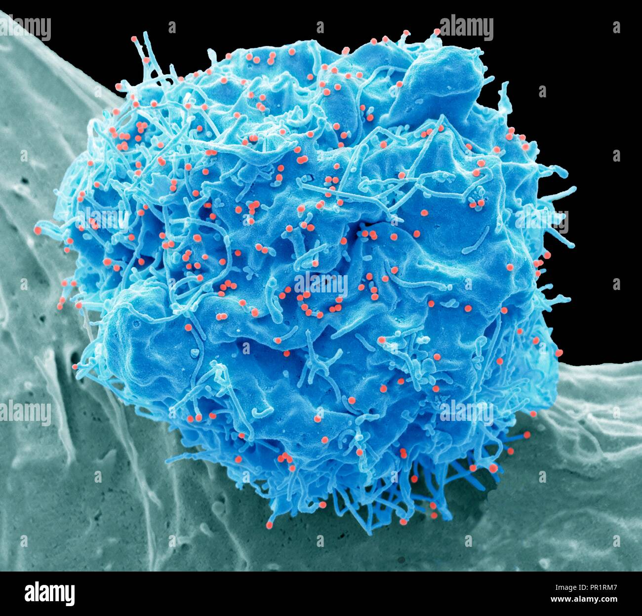
Ultrastructural analysis of SARS-CoV-2 interactions with the host cell via  high resolution scanning electron microscopy | Scientific Reports
Ultrastructural analysis of SARS-CoV-2 interactions with the host cell via high resolution scanning electron microscopy | Scientific Reports

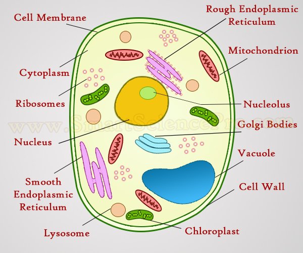
Structure of Animal Cell and Plant Cell Under Microscope + Diagrams

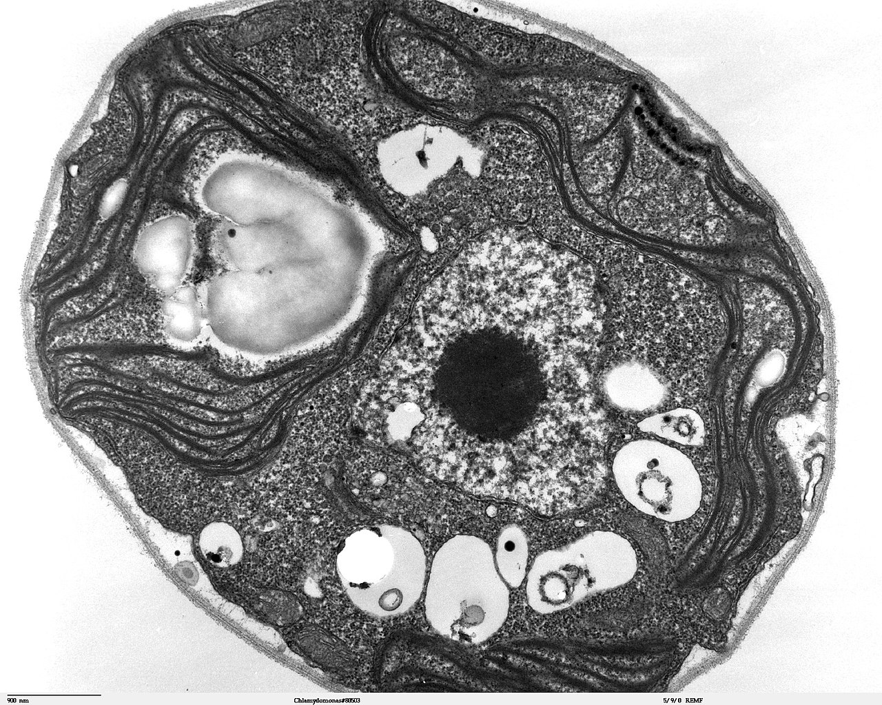
File:Chlamydomonas TEM 07.jpg - Wikimedia Commons

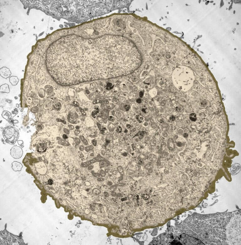
To peer inside a living cell | MIT News | Massachusetts Institute of  Technology
To peer inside a living cell | MIT News | Massachusetts Institute of Technology

1.2 Skill: Interpretation of electron micrographs - YouTube

323 Golgi Microscope Images, Stock Photos & Vectors | Shutterstock

New Microscopy Technique Shows Cells' 3-D Ultrastructure in New Detail |  HHMI
New Microscopy Technique Shows Cells' 3-D Ultrastructure in New Detail | HHMI

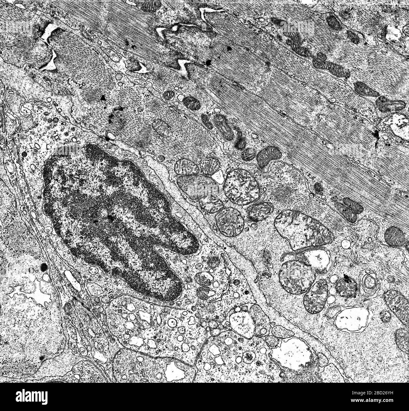
Electron microscopy human cell hi-res stock photography and images - Alamy

Cell nucleus and organelles under the electron microscope 50,000x Stock  Photo - Alamy
Cell nucleus and organelles under the electron microscope 50,000x Stock Photo - Alamy

Starch in chloroplasts vegetal cell under electron microscopy, Stock Photo,  Picture And Rights Managed Image. Pic. N88-3918251 | agefotostock
Starch in chloroplasts vegetal cell under electron microscopy, Stock Photo, Picture And Rights Managed Image. Pic. N88-3918251 | agefotostock

Amazing 27 Things Under The Microscope With Diagrams

Can people see eukaryotic cells under a scanning electron microscope? If  so, are there any images of that? - Quora
Can people see eukaryotic cells under a scanning electron microscope? If so, are there any images of that? - Quora
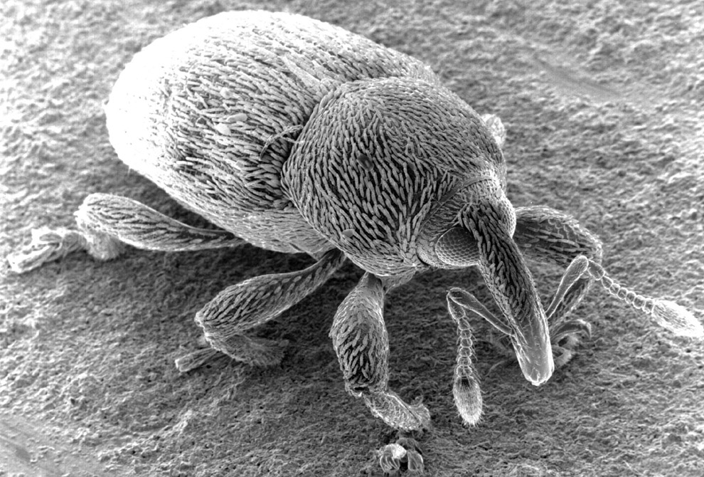
Peering into the micro world - Photos - The Big Picture - Boston.com

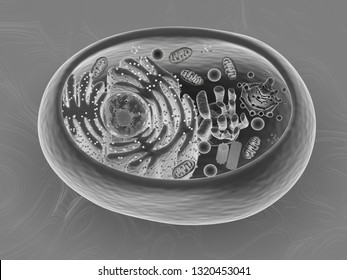
File:Anatomy and physiology of animals animal cell electron microscope.jpg  - Wikimedia Commons
File:Anatomy and physiology of animals animal cell electron microscope.jpg - Wikimedia Commons

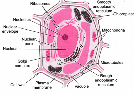
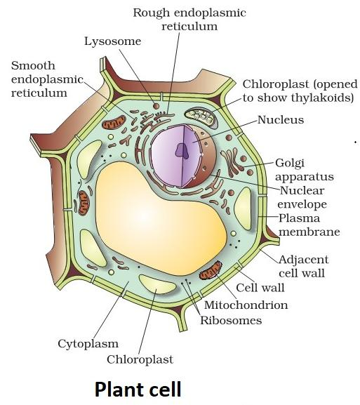
Illustrate only a plant cell as seen under electron microscope. How is it  different from animal cell? - CBSE Class 9 Science - Learn CBSE Forum
Illustrate only a plant cell as seen under electron microscope. How is it different from animal cell? - CBSE Class 9 Science - Learn CBSE Forum
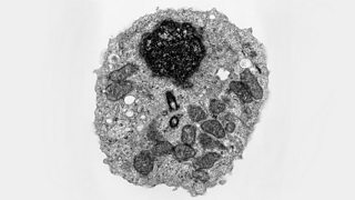
Electron microscopes - Cell structure - Edexcel - GCSE Combined Science  Revision - Edexcel - BBC Bitesize
Electron microscopes - Cell structure - Edexcel - GCSE Combined Science Revision - Edexcel - BBC Bitesize

Clinical Anatomy & Operative Surgery - Cancer cells under an electron  microscope | Facebook
Clinical Anatomy & Operative Surgery - Cancer cells under an electron microscope | Facebook

Cancer cells under an electron microscope : r/pics

Visita il nostro sito templedusavoir.org) | Electron microscope, Electron  microscope images, Microscopic photography
Visita il nostro sito templedusavoir.org) | Electron microscope, Electron microscope images, Microscopic photography
cell and organelles Dr.Jastrow's electron microscopic atlas

Illustrate only a plant cell seen under an electron microscope. How is it  different from animal cells?
Illustrate only a plant cell seen under an electron microscope. How is it different from animal cells?